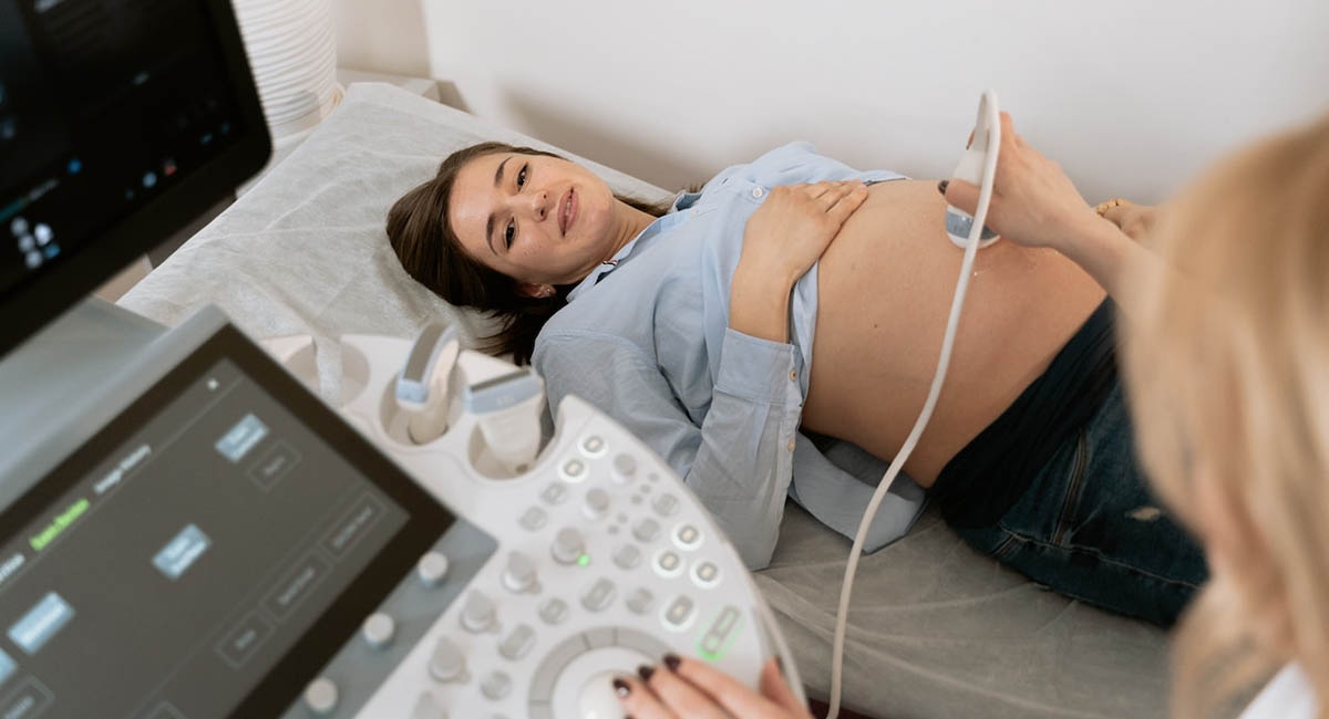
How Many Ultrasounds During Pregnancy
The use of ultrasound technology in ob gyn is one of the more controversial issues among expectant parents these days. But how many ultrasounds during pregnancy, and how often should you make? Let’s check that out.
Ultrasound Basics
If you are pregnant and if you are over the age of 18, an ultrasound is a great way to look at the fetus. Ultrasounds are performed in more than 90% of all pregnancies. They are used to confirm a pregnancy, determine gestational age, and detect fetal problems.
Types of Ultrasound
Several types of ultrasounds are used during pregnancy, depending on the situation. Diagnostic ultrasound is performed to see if the fetus has a problem. An abdominal ultrasound is a common diagnostic test involving a transvaginal abdomen scan. The doctor will use a wand with a microphone on end to bounce sound waves off of the inside of the womb. This creates an image of the baby.
Abdominal ultrasound exams help determine the best time for delivering your baby and are often used for non-risk screening. A screening ultrasound is usually used before 16 weeks gestation to determine if your baby is at risk for Down syndrome and other genetic disorders. Your doctor may suggest having a screening ultrasound at 11-13 weeks.
Why Is the Ultrasound Used?
If you are concerned about a possible fetal problem, ultrasound technology provides a detailed picture of your baby’s health and growth. It allows the ultrasound technician to make sure your baby is growing and developing normally and create an image of it. If you are concerned about your own health, such as gestational diabetes, you may also request that an ultrasound be performed. You can also check these motherhood tips, parenting, pregnancy & baby information to ensure a smooth and healthy pregnancy on the https://motherhoodtips.com/.
So here is what to expect from the ultrasound:
- Fetal Growth and Development – Ultrasounds are used to track the growth and development of your baby. This is done by determining the baby’s growth rate and its development;
- Fetal age – If the baby is older than 12 weeks gestation, the doctor will probably do an ultrasound to determine the baby’s gestational age. This information obtained during the early ultrasound is used to determine the week of the pregnancy;
- Detecting a problem – If the baby has a problem that requires a doctor’s attention, the ultrasound comes to the rescue; high frequency sound waves also allow to find out if there is a birth defect;
- Treating problems – Ultrasounds provide the best images of your baby. This allows your doctor to determine if there is a problem and, if there is, to find the best treatment options for the baby. What is more, ultrasound offers a rapid, non-invasive way to evaluate amniotic fluid;
- Risk vs. good health – Although doctors use ultrasounds to determine the fetus’s health, your health is also an important factor when deciding when and how often to have an ultrasound. If you are at risk for a condition that the ultrasound technology may not detect, you may request that the ultrasound be performed earlier. For example, if you have diabetes, you may want an ultrasound early in the pregnancy to closely monitor the baby. The early trimester ultrasound is especially important if your blood sugar levels are unstable;
- Fetal position – Your doctor will do an ultrasound to determine if your baby is head down or face down. This helps your doctor determine when the baby will be born because face-down babies usually arrive within 24 hours of the scan;
- Pregnancy risk – Many problems occur in the last trimester of pregnancy, which is why your doctor will perform an ultrasound after your 20th week to check on the baby. What is more, ectopic pregnancy can be easily detected thanks to the ultrasound;
- Non-Risk Screens – Your doctor may do a screening ultrasound before your 16th-week gestation. In this case, your doctor uses a wand with a microphone on end to bounce sound waves off of the inside of the womb. This creates an image of your baby and gives your doctor an idea of whether the baby is at risk for Down syndrome. Screening ultrasounds are used to find out if there are other problems that may indicate a high risk for Down syndrome. A screening ultrasound is used at 11-13 weeks gestation to determine if there is a problem with your baby’s heart, placenta, cervix, or any other part of the baby’s body. This is highly important for pregnant women who have already had a baby with Down syndrome or with a heart condition.
The First Ultrasound
Before you are pregnant, you will probably be tested for various conditions. Depending on the condition, you may want to schedule your first prenatal appointment for a diagnostic ultrasound. Some conditions can be detected through a screening test. Most ultrasounds are done to diagnose an abnormal condition in the fetus. This may involve the following tests:
- Amniocentesis – This test can be done around the 12th week of pregnancy. This test removes fluid from the uterus and tests for abnormalities. In the rare situation where a miscarriage is suspected, the doctor will perform an amniocentesis to check for problems;
- Non-risk screens – A screening ultrasound is used to determine if there are other problems that may indicate a high risk for Down syndrome. This is especially important for women who have already had a baby with Down syndrome or there were cases of a heart condition in the family history;
- Abdominal ultrasound – An abdominal ultrasound is used to detect and diagnose abdominal problems, such as an umbilical hernia or a bowel obstruction.
Most healthy women receive two ultrasound scans during pregnancy. In the ideal scenario, the first is in the first trimester to confirm the due date. In contrast, the second ultrasound is usually scheduled between 18 and 22 weeks to confirm normal anatomy and the sex of the baby. However, some additional ultrasounds may be necessary. And you should not worry since ultrasounds are safe.If you need any additional Ultrasounds you can always look at a private ultrasound.
Photo:MART PRODUCTION, Pexels


